Manual therapy

Manual therapy based on the Cyriax, DGMM and Dorn-Hock methods
Mainly used in the orthopaedic and traumatology sectors, but also indispensable in the treatment of medical and neurological disorders. The method enables functional diagnostics and the treatment of dysfunctions such as pain, hypo- and hypermobility, and of soft-tissue lesions affecting the locomotor system. Its aim is to maintain or re-establish normal function in the joints and all other functionally or structurally related locomotor tissue
Joint disorders
arising from arthralgia as a result of referred articular pain or indiscernible changes to joints.
Arthritis
following an injury involving damage to the joint capsule, sprain, luxation or subluxation.
Arthritis
following bacterial infection of joint; inflammatory rheumatic disorders; arthrosis; congenital joint deformities; metabolically-caused joint diseases such as gout and haemophilia; neoplastic arthropathies.
Joint capsular disorders
Typical pattern of restricted movement – the so-called "capsular pattern" or capsular sign – which reveals a different pattern of restricted movement in every joint. They occur in joints that are stabilized by muscular control. Joints that are sealed and supported solely by a capsule and ligaments do not exhibit a capsular pattern. If a capsular pattern should, however, be apparent, this indicates that the articular capsule is inflamed as in arthritis or arthrosis.
Ligament disorders
may arise when there is:
- acute ligamental injury, such as over-extension, partial rupture and total rupture;
- chronic injury and pain following over-exertion of joint, but in which the joint is functionally stable; or
- functional instability of joint, e.g. in a partially restored total rupture.
- Bursitis
Physiotherapy must not be used to attempt treatment of a traumatic, infectious or tumour-related inflammation of a bursa.
"Bursitis calcarea"
Deposition of calcareous matter through the bursa into the rotator cuff, only very occasionally in the bursa itself.
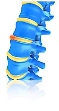
Protrusion of the fibrous ring (Anulus fibrosus). Develops gradually, pain is localised. No loss of neurological function, but patient typically adopts a temporary posture to relieve discomfort.
Prolapse:
Disc no longer intact, e.g. due to rupturing of the anulus fibrosus and protrusion of the pulpy core. Loss of neurological function, reduced sensitivity and loss of strength, reflex response lower (hyporeflexia). Onset is sudden. Symptoms include parasthaesia and referred pain. Patient adopts a permanent relieving posture.
Disorders of the facet joints:
Non-specific pain caused by impeded movement of vertebral joints. The impediment is due to damage to the vertebral discs, which reduces the inter-disc spacing and increases the compressive loading on the vertebral joints.
Referred pain in dermatome:
Referred pain (pain felt at some distance from its cause) can result from disorders of the:
- Joints (capsules, ligaments, bones)
- tendomyotic system (tendon, tendon insertion, muscle-tendon junction, muscle belly)
- internal organs.
When treating disorders of the locomotor system, the aim is to clearly identify the affected areas. This requires that the affected areas be distinguished clearly from those that could give rise to the same clinical characteristics.
McKenzie
Different syndromes are defined, which can be the basis for painful conditions and restricted movement of the postural and musculoskeletal system and are classified according to the diagnostic scheme developed by Robin McKenzie.
Postural syndrome:
Posture-related pain can be caused by all pain-sensitive structures of the holding apparatus (e.g. spine) due to overstretching of normal, healthy structures.
Dysfunction syndrome:
Restriction of movement caused by adaptively shortened structures. The same mechanism applies here, except that the pain is the result of the stretching of adaptively shortened or abnormal tissue.
Derangement syndrome:
Most often in patients between 20 and 55 years of age, M> F. The pain in derangement syndrome is explained by a shift or changed position of joint structures, which leads to mechanical deformation of pain-sensitive structures. This results, for. B. a changed position of the joint surfaces to each other, which can manifest itself clinically in incorrect posture and deviation from the normal trajectory. Different derangement syndromes are distinguished depending on the location and radiation of the pain.
The assignment
the three categories are based on the findings. It is based on the change in symptoms after repeated movements during tests and treatment.
 DE
DE
 FR
FR